CT can be the quick first look
Fast, widely available and often cheaper. Useful for urgent bleeding, large stroke, hydrocephalus, tumor or subdural collection.
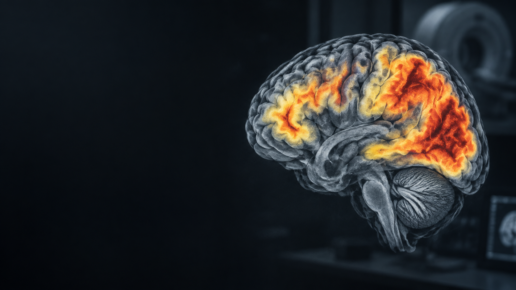
CT · MRI · PET
Scans can reveal structure, injury, blood-vessel disease, metabolism and selected protein targets. None should be read alone.
See a possible pathwayA possible pathway
The sequence varies by urgency, availability, cost and the clinical question. MRI may be the better first test when detailed dementia assessment is already the goal.
If there has never been a brain scan, or the last scan was some time ago and symptoms have changed, a clinician may recommend current structural imaging to establish a baseline and check for a new cause.
Fast, widely available and often cheaper. Useful for urgent bleeding, large stroke, hydrocephalus, tumor or subdural collection.
Better for hippocampal and regional volume loss, small infarcts, white-matter disease, microbleeds and traumatic injury. Some people go straight to MRI.
FDG-PET examines metabolism. Amyloid and tau PET target specific proteins when specialist assessment needs additional evidence.
Important: CT first does not always save a second scan. If MRI is likely to be needed anyway and is suitable, going directly to MRI may be more efficient.
Compare scans
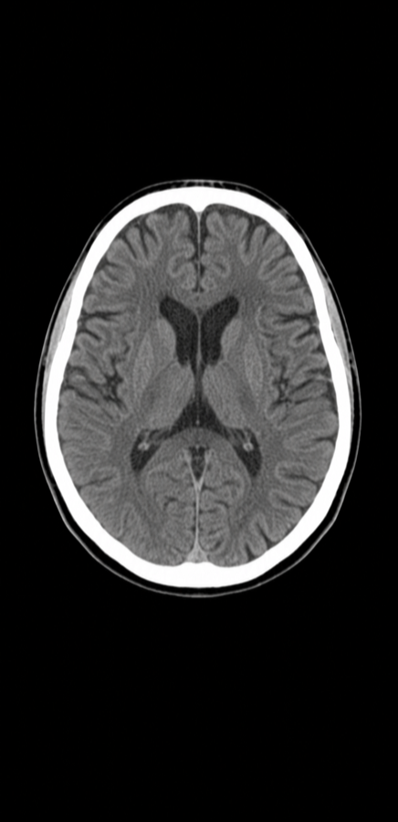 Structural · X-ray
CT can identify bleeding, a large stroke, tumor, hydrocephalus, subdural collection and major tissue loss.
Illustrative teaching images show how modalities differ visually. They are not patient scans and cannot be used for diagnosis.
Beyond imaging
Routine blood tests may identify reversible or contributing problems such as thyroid disease, vitamin deficiency, infection, organ dysfunction or metabolic disturbance. Specialist biomarker blood tests can measure proteins associated with Alzheimer's pathology, but still require clinical interpretation.
Explore Alzheimer's testsPattern atlas
Illustrative teaching patterns, not real patient records. Appearance varies with stage, technique and coexisting disease.
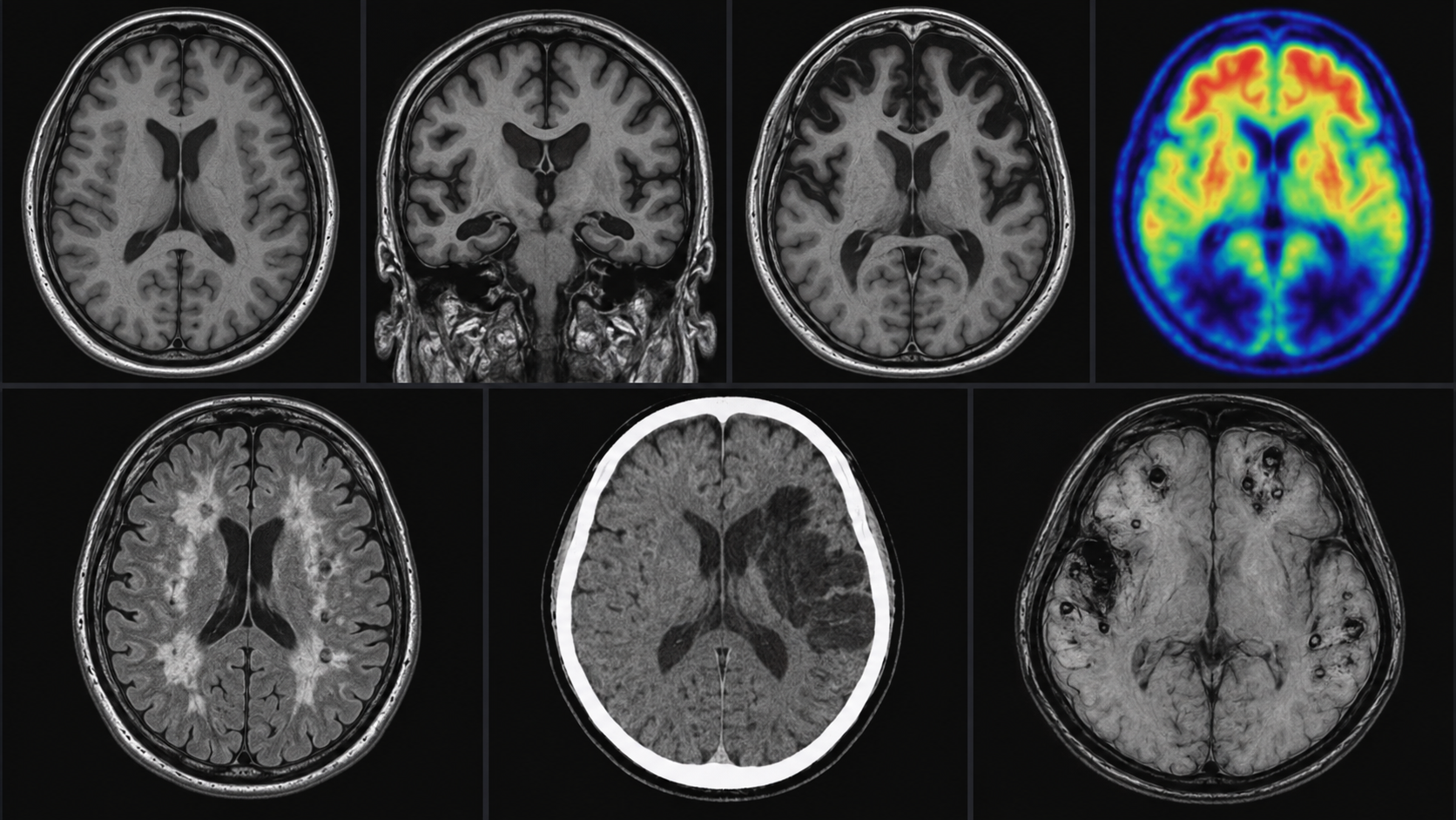
Some volume loss and white-matter change can occur. Distribution and severity matter.
Medial temporal and later temporoparietal volume loss; selected PET assesses metabolism, amyloid or tau.
Frontal and anterior temporal volume loss or reduced metabolism, sometimes asymmetric.
Structural MRI may be nonspecific. Selected functional or dopamine-transporter imaging may support assessment.
Infarcts, lacunes, microbleeds and extensive white-matter disease may support a vascular contribution.
CT is central acutely; MRI can define the location, age and extent of ischemic injury.
CT detects many acute fractures and bleeds; MRI may reveal contusions and diffuse axonal injury.
Do not miss
Progressive confusion or personality change is not always a primary dementia. CT or MRI may reveal another cause requiring a different response.
An infiltrating malignant brain tumor can cause cognitive, personality, language or neurological change. MRI with contrast usually defines it better than CT.
A slow-growing meningioma may compress frontal or other brain regions and present subtly. Imaging shows its site, mass effect and surrounding edema.
Weeks after a minor fall, particularly in older people or those using blood thinners, a chronic subdural can cause worsening cognition, gait or weakness. The fall may have been unwitnessed or forgotten, and there may be no scalp bruise or visible injury. An anonymous Anonamed profile may help identify medicines and conditions without exposing identity or contacts on a locked phone. Named emergency contacts may be available separately through Apple Medical ID or a clearly marked wallet note or card. A wallet card can also carry the anonymous Anonamed QR code for access to the medical profile. For someone with early dementia who remains independent, this can be combined with a separate contact card, itinerary and check-in plan when going out alone or travelling with a group. Anonamed is not a tracker and does not identify the person. An accessible advance care directive may communicate treatment preferences and limitations. CT often detects a subdural quickly.
Read the whole picture
A normal scan does not end the assessment. Early disease may precede obvious structural change.
An abnormal scan is not automatically dementia. Age, vascular risks and previous injury alter appearance.
Amyloid is not the same as symptoms. A positive scan must be interpreted in context.
Evidence